Dissection
A dissection is a tear on the inside of a blood vessel. It can lead to both a cerebral hemorrhage and a cerebral infarction. The arterial wall consists of three layers, from the inside out: the intima, the media and the adventitia.
Blood first seeps between the inner wall and the outer wall of the blood vessel. This can cause a separate channel through which blood flows. This can transform into a balloon. This balloon can become so large that it can pinch nerves and other blood vessels. This can lead to a cerebral infarction.
If the balloon bursts or the blood vessel ruptures further, it can lead to a cerebral hemorrhage.
A dissection leading to a cerebral infarction or cerebral hemorrhage most commonly occurs in the internal carotid artery, the vertebral artery or the basal cerebral artery (basilar artery).
The latter artery is responsible for 20% of the brain's blood supply and runs at the bottom of the brain, the base. The middle image below shows how the inside of the blood vessel ruptures and a 'false' aneurysm (balloon in the vessel wall) is created. The resulting balloon may lead to an infarction.
Risk factors for a vessel wall rupture: high blood pressure, high cholesterol in the blood, smoking, respiratory infection of the upper respiratory tract, an accident to the neck area or (hereditary) connective tissue disorders and hereditary or congenital disorders. Also an Autosomal Dominant Polycystic Kidney Disease (ADPKD) can lead to a dissection. If such a vessel wall rupture occurs, it is an acute and life-threatening situation.
If several blood relatives within one family have had a CVA / stroke due to a vascular abnormality, this may be a reason to conduct family research.
There are English Facebook groups for people with carotid artery dissection:
A public group: https://www.facebook.com/groups/47265659349/?hc_location=ufi
A private group: https://www.facebook.com/groups/606107169461087/?hc_location=ufi
Images below:
1) the internal carotid artery (internal carotid artery):


by Heusser
3) basilar artery, the artery drawn in the middle:
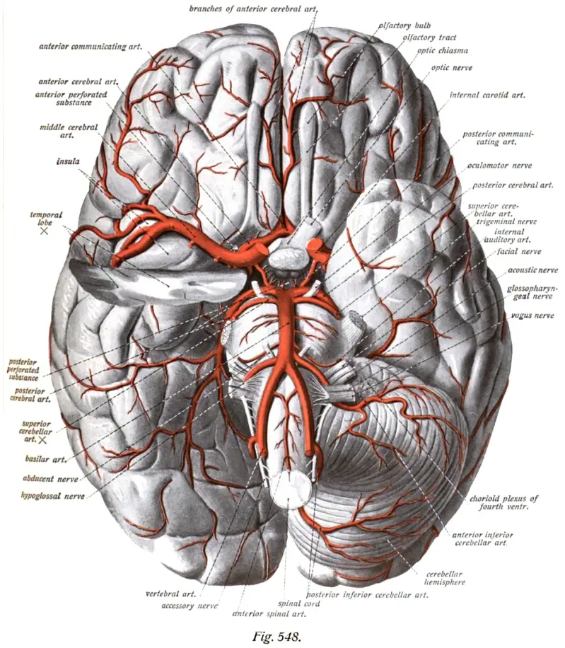
Image from Wikimedia
Resources
Brandt, T. (2000). Cervical Artery Dissection: Update and New Results of Research of the Pathogenesis. Cerebrovascular Diseases, 10(4), 5–8. https://doi.org/10.1159/000047583
Cline, D. M., Ma, O. J., Meckler, G. D., Tintinalli, J. E., Stapczynski, J. S., & Yealy, D. (2015). Tintinalli's Emergency Medicine: A Comprehensive Study Guide, 8th edition (8e ed.). New York, V.S: MacGraw Hill.
Eyskens, E., Feenstra, L., Meinders, A. E., Vandenbroucke, J. P., & Van Weel, C. (1997). Codex Medicus (10e ed.). Maarssen, Nederland: Elsevier Gezondheidszorg.
Haggstrom, M. (2007, 6 oktober). File:External carotid artery.png - Wikimedia Commons. Consulted on 20 december 2016, van https://commons.wikimedia.org/w/index.php?curid=2867505
Häggström, M. (2014). Medical gallery of Mikael Häggström 2014. WikiJournal of Medicine, 1(2). https://doi.org/10.15347/wjm/2014.008
Hersenletsel-uitleg team
Kuks, J. B. M., Snoek, J. W., Oosterhuis, H. G. J. H., & Fock, J. M. (2003). Klinische neurologie (15e ed.). Houten, Nederland: Bohn Stafleu van Loghum.
Veltkamp, R., Veltkamp, C., Hartmann, M., Schnffeldt-Varas, P., & Schwaninger, M. (2004). Symptomatische Dissektion der Arteria carotis interna. Der Nervenarzt, 75(2), 149–152.https://link.springer.com/article/10.1007/s00115-003-1644-9
and by dr Johannes Sobotta, public domain wikiwand.com