Visual consequences of brain injury
No eye damage but brain damage
You look with your eyes and you see with your brain. The brain and eyes are connected by optic nerves.
The brain plays a very important role in our ability to see everything clearly. Damage to the brain can therefore lead to visual complaints.
After brain injury, the brain cannot always make a good picture of what you see.
The brain then needs more effort and more time to properly interpret or process what you see. If the processing of visual stimuli in the brain is reduced or disrupted, this is called a visual processing disorder.
There may also be complaints because one of the three specific cranial nerves does not control the eye muscles or does not control them properly due to the brain injury.
A large brain network processes the images presented by the eyes. That's more than just the occipital “visual” cerebral cortex in the back of your mind.
Damage caused by non-congenital brain damage, such as a stroke or an accident or abnormal development can lead to a variety of visual disturbances.
For example:
- no longer being able to read after sustaining brain damage
- visual overstimulation
- inability to recognize objects or familiar people
- only seeing one thing at a time
- double vision
- part of the field of view may disappear,
- spots in the field of vision
- varying sharpness of vision (think of CVI!)
- refractive problems (not being able to see clearly)
- information from one eye can be processed faster than information from the other eye
- delayed information processing of the visual system. This means that someone can continue to see a tree even though the tree has long been out of sight, for example. See 'locking image' below.
- fixation disparity problems, where the eyes do not work together properly
- due to inability to fixate, reading can be difficult or impossible
- light hypersensitivity, sensitivity to bright light or sunlight (photophobia).
- visual overstimulation.
There is nothing wrong with the eyes, but there is damage to the visual brain areas or the nerve pathways leading to them.
Certain vision problems can also be a result of loss of balance, damage to balance or certain balance diseases.
Vision problems can lead to severe fatigue, headaches, pain around/in the eye and feeling 'seasick or drunk'. People may feel like their eyes are running wild.
Quote 1
Due to my cerebral infarction I miss a quarter of my vision.
In the beginning, my brain filled in that quarter itself and so I saw strange things in my left field of vision, the entire left side also moved, while the right remained still (think of faces, paintings, roads, sidewalks).
That disappeared over time.
I couldn't read printed text at the time (fortunately I can now).
My vision remains cloudy as if I have “fog” in my eyes.
The further away I can look, the clearer it becomes.
Quote 2
I had problems with what I call 'image freeze'.
When this is fixed, it is no longer possible to see another image,
so for example you see a tree and if you continue walking you continue to see that tree.
Sometimes this phenomenon precedes injury due to a reduced amount of oxygen in the blood.
Complaints may include:
- Not being able to read
- Visual overstimulation, including for light and movement
- Double vision or muscle weakness of eye muscles (diplopia) (double images, which are seen as separate by the brain, dance together)
- Blepharospasm or eyelid spasm. It causes spontaneous contractions of the eyelid muscles, forcing a person to focus again and again.
- No depth perception
- Trembling googly eyes (nystagmus)
- Visual field loss of seeing half (hemianopsia) or a quarter (quadrantopia), seeing parts or seeing only the edges (concentric limitation) or seeing the bottom half.
(Left-sided brain damage leads to loss of the right half of the visual field, and damage at the top leads to loss of visual field at the bottom.)
- No longer able to see colors
- Only being able to see one thing at a time.
- Photohypersensitivity, sensitivity to bright light or sunlight (photophobia)
- Blurred, unable to see clearly, despite good glasses correction
- Varying sharpness of vision (think of CVI!)
- Refractive error (inability to see clearly)
- Information from one eye is processed faster than from the other eye
- 'Freeze image'. When you freeze it, it is no longer possible to see another image, so for example you see a tree and as you continue walking you continue to see that tree. Fragmented vision due to delayed information processing. Sometimes this phenomenon precedes injury due to a reduced amount of oxygen in the blood.
- Difficulty estimating distances correctly. (think of CVI!)
- Not able to recognize objects, read more on our page on agnosia
- No longer able to recognize faces / facial expressions, read more on our page on prosopagnosia
- Impaired visual memory
- Seeing spots in the image (scotoma) or 'visual snow' as if noise or flashes of light are seen in the image
- Impaired spatial judgment (orientation) and movement in a space in relation to people and objects
- Difficulty estimating distances or speed, but also incorrectly estimating distance when taking steps / stairs
- Fixation disparity problems, where the eyes do not work together properly.
- Inability to fixate can make reading difficult or impossible (possibly possible through appropriate reading, Daisy player for audio books).
- Convergence insufficiency, in which you cannot see at close range because the eye muscles do not turn properly towards the nose.
- People with MS may have damaged optic nerves and have Uhthoff's syndrome. This means that complaints temporarily worsen under the influence of fatigue or heat such as warm weather, increased body temperature, for example due to a warm bath or physical exertion. The vision problems then increase in the form of blurred vision or double vision until cooling occurs.
- Visual overstimulation is a result of damage in the brain, whereby a person consciously perceives so many details that it is simply too much for the brain. The light may be perceived as too bright. The light reflection is so striking that someone cannot close themselves off to it.
Seeing letters of text that are too close together or seeing a screen can become sickening in a very short time. With visual overstimulation, someone can consciously and amplify so many details that it is simply too much for the brain.
Seeing busy patterns or colors, a multitude of cozy things in the house, or seeing movements can also lead to visual overstimulation. Visual overstimulation can cause someone to lose balance.
The type of complaints a person has depends greatly on the location of the damage in the brain.
Brain damage rarely involves a visual disturbance alone, see our other pages about consequences.
Practical consequences
The visual complaints can cause limitations in:
- reading, watching TV, computer work
- in traffic, because it is more difficult to find your way
- recognizing objects or faces even of famous people, "I didn't even recognize my son" Read more..
- performing household tasks
- grabbing objects Read more..
Where is the injury?
Visual complaints due to brain injury can occur in several areas depending on the symptoms:
- Simultaneous agnosia in parietal lobe: yellow in the image below
- Associative agnosia usually in the left hemisphere, in
occipital lobe, border temporal lobe: green/red in the image below:

- Prosopagnosia, no longer being able to recognize faces, gyrus fusiformis in the picture below:

Most visual complaints arise after damage to the occipital lobe, including apperceptive agnosia, which usually occurs in the right half of the occipital lobe: red in the image below:

Eye muscles and three cranial nerves
Each eye has six muscles. Cooperation between the muscles of the eyes allows the eyes to see in a coordinated manner and in this way double vision is prevented.
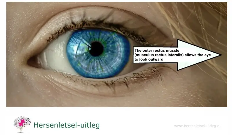
Brain damage can cause an eye muscle to fail. Then the muscle cannot cooperate with other muscles. As a result, the images of the two eyes do not coincide.
Each eye has six eye muscles that are controlled by different cranial nerves from the brain: III, IV and VI.
Normally this allows the eye to turn in all directions.
Looking inward is called adduction. Looking outside is called
abduction. Looking up is called elevation and looking down is called depression. Turning inwards is called intorsion. Turning outwards is called extortion.
If one of these three cranial nerves transmits less or no information, one or more eye muscles do not work properly. There is then an inability to look at a certain side with both eyes; gaze paralysis or gaze palsy. Double images and other complaints may arise. A gaze palsy can also occur due to damage in one of the gaze centers. We will discuss this further on this page.
Drop-down menu:
Gaze centers
The gaze centers are areas in the brain that ensure that the eyes can be controlled simultaneously to produce parallel (conjugate)
eye movements.
The eye movements can be divided into:
- Saccades. These are the horizontal movements of the eyes to be able to focus very quickly on something. Both reflexively and consciously looking at something.
- Tracking movements. These are the eye movements to follow a moving object.
Gaze centers in the cerebral cortex:

- the prefrontal cortex in the forehead: frontal gaze center, for simultaneously controlling horizontal eye movements and consciously controlled search movements (Brodmann area 8)
- the parietal cortex in the parietal lobe for the saccades.
- the parieto-temporo occipital cortex (PTO) for unconscious fixation and tracking movements. This is also where visual-spatial recognition takes place and is important for the accurate location of what a person sees.
Parts of the gaze centers are located in the parietal lobe, temporal lobe and occipital lobe, hence the abbreviation PTO
Gaze centers in the brain stem:
- midbrain (mesencephalon): mesencephalic gaze center, for vertical eye movements
- bridge of varol (pons): pontine gaze center for horizontal eye movements and coordinating which cranial nerves are controlled
Gaze paralysis / gaze palsy
If damage has occurred in the vision centers due to brain damage, the eyes cannot look in one direction simultaneously. This is called gaze paralysis or gaze palsy.
- Vertical Gaze Palsy: A person with this condition cannot look down/up with both eyes at the same time. The brain damage then occurs in the midbrain (mesencephalon).
- Horizontal Gaze Palsy: A person with this condition cannot look to the side with both eyes at the same time. In diseases of the cerebral cortex of the cerebral hemispheres (cortical gaze palsy), horizontal gaze palsy may occur. The eye muscles themselves are undamaged.
Horizontal gaze paralysis can also occur with injuries to the pons.
See this website for more information.
Some quotes from people with gaze palsy:
- Due to traumatic brain injury, I have: double vision, eye strain, short focus, difficulty with moving images, strabismus, limited load tolerance, alternating between tasks near and far. (Melissa)
- Bacterial meningitis caused me to see double. The sixth nerve appeared to be affected. This caused my right eye to turn inwards. I wore an eye mask for months and my eye eventually healed itself after about 6 months. When I get tired I start to see double again, but my eye is miraculously straight again. (Marike)
- After a traumatic brain injury, I suffered from double vision and could not tolerate my glasses, although when checked by the ophthalmologist my glasses fit my eyes properly. (Frank)
Visual field loss
The visual field is what you can see with one eye. Visual field is everything you can see when you look straight ahead with both eyes.
With both eyes you see more than with one eye alone. A loss in the visual field or field of vision is like a blind spot. A large part of the visual field is lost.
This is not always noticed immediately, because the other eye can compensate a lot.
The field of vision contains 95 degrees of vision on both sides of the head. Each eye has 60 degrees of vision.
In the image below you can see that the field of vision of the right eye is shown in green and that of the left eye in orange. You can see that there is overlap when you look straight ahead (represented by both the green and orange lines).
That is why loss in the central field of vision, i.e. when looking straight ahead, is not noticed immediately.

Convergence insufficiency
When the eyes do not turn towards the nose the cause is often a combination of reduced cooperation and reduced functioning of the inner eye muscles. You cannot look at the tip of your nose.
The eye problems mentioned are called convergence disorder or convergence insufficiency. The movement of the eye muscles towards the nose is called convergence.
Convergence allows you to see at a short distance. This is important for work at a computer, reading, writing and for many daily tasks.
Complaints may include:
- headache (above the eyes or on the forehead). The eyes become tired more quickly due to strained close-up viewing
- blurred vision of what is close
- double vision of what is close
- seeing moving or dancing letters when reading
Search for help
The problem of double vision, which causes visual overstimulation, can possibly be reduced by an ophthalmologist or by an optometrist with experience in functional neurology and knowledge of brain damage, depending on the complaints.
Sometimes a chiropractor with experience in functional neurology can also provide help.
Resources
The Dutch page on which this page is based was created by Hersenletsel-uitleg.nl in collaboration with Koninklijke Visio and Lievenberg Hospital Neurology for the tips.
Dr. Ben van Cranenburgh, The hierarchical structure of the nervous system, Bohn Stafleu van Loghum 2020 Published in: Neurowetenschappen