Mesenchephalon - Midbrain
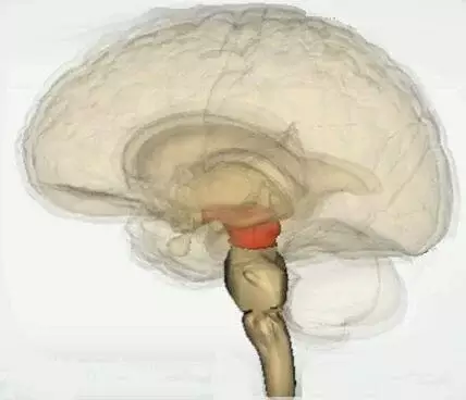
Physiologically, the brain can be divided into three parts:
- Forebrain
- Midbrain
- Hindbrain
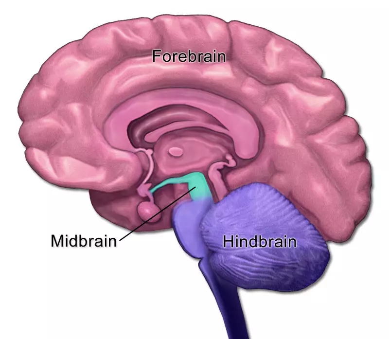
The midbrain is the upper part of the brainstem
The midbrain is located below the cerebral cortex and above the hindbrain, near the center of the brain.
Hence the name midbrain.
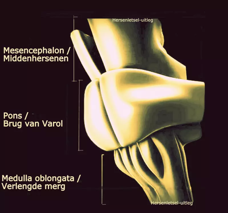
It is about 2.5 centimeters long and has a central location in the cranial cavity. We distinguish the brainstem into three parts.
The upper part is formed by the midbrain.
The lower parts belong to the hindbrain.
From top to bottom:
- Midbrain (mesencephalon),
- Pons (Bridge of Varol),
- Medulla oblongata.
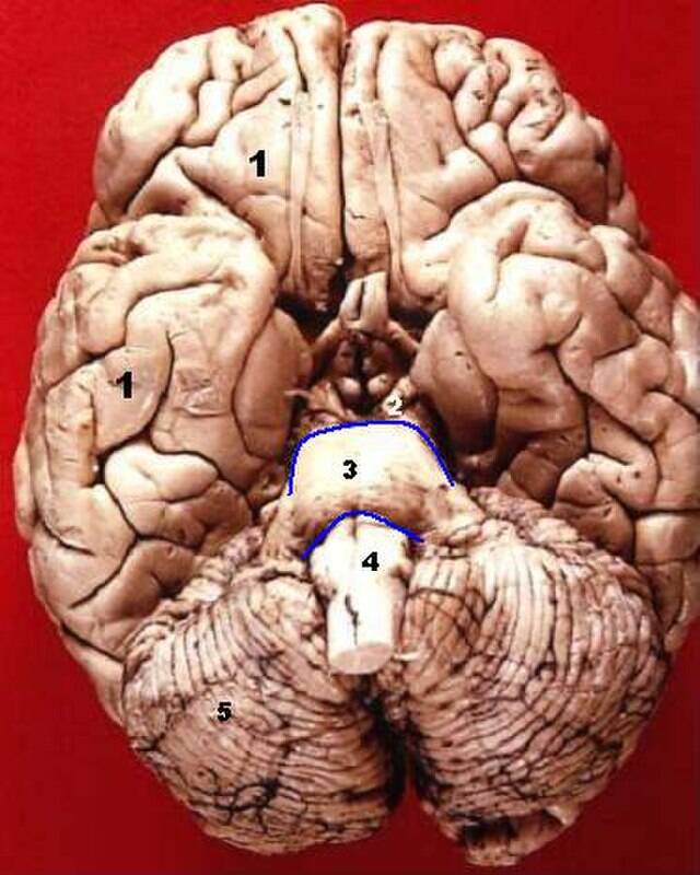
The image below shows the front of the three parts of the brainstem.
Through the midbrain are a number of nerve pathways that connect the cerebrum to the cerebellum and to the nerve bundles that form the reticular formation and other structures of the hindbrain.
The midbrain consists of:
-
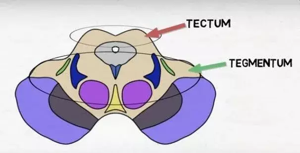
Tectum mesencephali or four-hilled plate / lamina quadrigemina. Tectum means 'roof'; roof of the midbrain. The function of the tectum mainly includes visual integration, eye movements and auditory functions. The tectum has four bulges on the dorsal side:
-
Two superior colliculi where information from the eyes is processed and where a reflex is generated in case necessary
-
Two inferior colliculi where information from the ears is processed and where a reflex is generated in case necessary
-
-
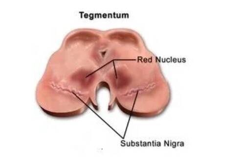
Tegmentum mesencephali which literally means the 'rug', the bottom of the midbrain. It inhibits, among other things, involuntary movements. The tegmentum includes part of the reticular formation. Some large collections of cells that fall under this 'formatio reticularis' are:
-
Red Nucleus (Red Nucleus (RN) or nuclei ruber) an important switching nucleus of the extrapyramidal motor system. This is where muscle tension and posture are regulated.
-
Black nuclei (Substantia Nigra) where dopamine-producing nerve cells are located.
-
- Aqueduct of Sylvius or cerebral aqueduct, a cavity filled with cerebrospinal fluid that connects the third and fourth cerebral cavities or chambers (ventricle)
- Cerebral peduncle (Crus cerebri; Crus means 'pillar' and cerebri means 'brain' = 'pillars of the brain') that contains motor nerve pathways that run from the cerebral cortex to the pons in the brain stem and the neck and back
- Cranial nerves (cranial nerves)
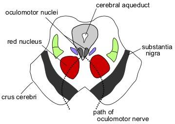
Image above: section through the middle of the mesencephalon at the level of the superior colliculus (upper bulge). The cerebral aqueduct is located on the back. The image below also shows a cross-section.
The image below shows the brain from below. The midbrain is shown with a white number '2'.

Function
The midbrain processes information from the ears and eyes and uses this information to make targeted movements.
Example: imagine you hear a car approaching. That signal is carried to the midbrain, which then activates the muscles of the neck to turn toward where the sound is coming from. In this way, the midbrain helps to orient to sound.
Another example: If you see a large black spider crawling downwards somewhere in a remote corner of your field of vision, a reflex follows that you don't think through... but all your muscles tense and you can get away.
The midbrain is involved in:
- The regulation of motor (movement) functions. In particular, the red nucleus (nucleus ruber) and the black nuclei (substantia nigra) are involved in the control of body movement,
- The regulation of muscle tension in different sleep states,
- Tactile information processing,
- Visual reflexes as a kind of relay station. It regulates the reflexes to acute changes in ambient light, such as the pupillary light reflex. It also controls the reflex to keep something centrally in view when you look and are moved: the optokinetic reflex (combination of smooth eye tracking movements and jumps/saccades). It is the coordination center for vertical eye movements.
- Auditory reflexes as a kind of relay station,
- Hearing,
- Waking and sleeping,
- Oral proprioception; the ability to perceive parts of the mouth. In particular, it prevents you from chewing your teeth on pieces of bone or hard stones.
Damage
Damage to the midbrain can affect all of the functions we just listed.
- Damage in regulation of sensory functions,
- Damage in regulation of motor functions,
- Damage in visual and auditory reflexes,
- Damage in eye movements; vertical gaze palsy.
With vertical gaze palsy, a person cannot look up or down with both eyes at the same time.
There is damage in the dorsal part of the midbrain (dorsal mesencephalon).
This is caused, for example, by a tumor or hydrocephalus.
Damage to the midbrain can cause various syndromes, with symptoms depending on the location in the midbrain:
- Benedict's syndrome
- Claude's syndrome
- Parinaud's syndrome
- Weber's syndrome
Because so many reflex actions are regulated in the midbrain, it is to be expected that disruption of defense responses may occur in the event of damage.
The reticular formation

The reticular formation (formatio reticularis or 'the reticular core') is a complex network of neurons. It consists of gray matter, which occupies space between white fiber tracts and brainstem nuclei. It runs through the midbrain and hindbrain and has connections to numerous areas in the cerebrum.
It is responsible for: the coordination of autonomic functions such as breathing, blood pressure, heart rate and pain perception, the level of consciousness, the sleep-wake rhythm, the regulation of the activation state of the nervous system. It is therefore called the control room of consciousness.
The reticular formation receives stimuli from outside the body via the senses and from the body via: propriosensors (registration of movement and posture in the vestibular organ, joints, tendons and muscles) nocisensors (registration of pain) interosensors (registration of the situation of the organs).
It helps the brain to 'screen' information/stimuli. They filter the unimportant information so that the person can focus on the important information that has the highest priority.
For example, if you look at a part of a city, with the sky above it, and at the bottom left of your field of vision a lawn and trees, a little in front of it on the left a group of people and on the right cars approaching, then the reticular formation will help the brain not to become overloaded with stimuli.
It will block the sky and the lawn, the thousands of tree leaves moving, from becoming aware. It will consciously transmit the moving cars and the group of people and some sounds.
As a result, later on you will only remember certain parts and not the tree leaves, for example, because the reticular formation has not passed on that information to be processed.
This is also the trick that illusionists use with illusions. They consciously use what people focus their attention on. The reticular formation does pass on important warnings to the higher brain areas to be alert. The reticular formation helps to be alert and alert, it regulates wakefulness and contributes to muscle tension necessary to take action.
Three important circuits can be distinguished in the reticular formation: reticulospinal circuit (motor functions such as muscle tension (muscle tone), reflexes and autonomic functions such as blood pressure, respiration and heart rate) reticulothalamocortical circuit (activation of the cerebral cortex / arousal) extrathalamic cortical circuit (activation of the cerebral cortex / arousal, in particular strengthening or weakening the excitability of the cerebral cortex).
RAS
The reticular formation contains a number of nerve nuclei that act as an alarm system: the reticular activating system (RAS).
The RAS has ascending (afferent) pathways (ARAS) to the cerebral cortex (cortex cerebri) and descending pathways to the spinal cord. The RAS spreads from the reticular formation in all directions of the brain, through the thalamus and to the spinal cord.
These nuclei are responsible for regulating wakefulness and arousal and stages of sleepiness and sleep-wake transitions. The sleep disorder narcolepsy is associated with the reticular activating system. People who suffer from narcolepsy can experience uncontrollable bouts of drowsiness at any time of the day. Conversely, the RAS can also ensure supreme concentration and can therefore ensure that people can deliver top performance.
Black core / Substantia Nigra (SN)
The black nucleus or substantia nigra is located in the anterior part of the mesencephalon and supplies the basal ganglia with dopamine. It is a motor nucleus. Despite its location in the mesencephalon, it is considered part of basal ganglia in function. The black nucleus is involved in voluntary movements, orientation in space, learning, mood regulation, and activity related to the brain's reward circuitry.
The substantia Nigra consists of:
- The pars compacta (SNc) on the dorsal side with melanin-filled neurons. Melanin is the dark color (pigment) that gives the black core its name. It supplies the striatum with dopamine and is very important for transmitting signals from the basal ganglia to the thalamus. It is involved in motor control and coordination. The loss of dopamine neurons in this part (SNc) appears to be responsible for the development of Parkinson's disease and some other parkinsonian syndromes.Dopamine is a signaling substance (neurotransmitter) that is related to movement disorders, psychiatric disorders and behavioral problems. See the chapter below.
- The pars reticulata (SNr) on the ventral side. SNr is important in the regulation of eye movements.
Dopamine deficiency or Dopamine excess
The dopamine-producing nerve cells are located in the black nuclei (substantia nigra) in the mesencephali tegmentum. In the upper part of the brain stem.
Dopamine is a signaling substance (neurotransmitter) that is related to movement disorders, psychiatric disorders and behavioral problems.
Together with serotonin and norepinephrine, dopamine ensures fine motor skills, coordination and concentration.
Addiction mechanisms
If we just look at the function of dopamine, which is sometimes called 'the happiness hormone', we see that dopamine is secreted at the brain level when receiving a reward, after effort, exercise, sports, sex, food, listening to pleasant music or when using certain medications, but also with nicotine or drugs. Overactive dopamine pathways can predispose a person to addictions.
Being able to make movements
Parkinson's disease is related to the death of nerve cells in the black nuclei, the substantia nigra. This causes a deficiency of dopamine. Dopamine also helps the body to make movements. Other diseases involving disorders of the musculoskeletal system, such as Huntington's disease, are also associated with reduced dopamine release.
Behavioral problems and neuropsychiatry
Disruption of dopamine release can also cause all kinds of mental complaints such as loss of concentration, nervousness, depression, lethargy, but also obsessive compulsive behavior (OCD) and ADHD. Dopamine keeps you motivated.
Lack of motivation, sleep deprivation, digestive problems, constipation and many more complaints are also associated with dopamine deficiency.
The opposite is also possible: overactive dopamine pathways can lead to neuropsychiatric disorders, such as schizophrenia.
This complex signaling substance has many undiscovered functions.
Lesion in the red core
When the red core is damaged, the following complaints may occur:
- Intention tremor. An intention tremor is the shaking of the limbs during a purposeful movement. It can be most easily observed in the hands, for example when gripping an object.
- Reduced muscle tone or even paralysis on the other side or half of the body (contralateral side).
- Specific movements:
- Choreic movement: involuntary, sudden, rapid, irregular movements that may occur in the arms, legs, face, trunk and neck both at rest and when moving.
- Athetotic movements: slow, uncontrolled, worm-like movements of the arms and legs.
Parinaud's syndrome
Parinaud's syndrome is an inability to move the eyes up and down.
Synonyms: Dorsal midbrain syndrome, Pretectal syndrome, Sylvian aqueduct syndrome, Koerber-Salus-Elschnig syndrome.
Parinaud's syndrome is divided into three complaints:
conjugated upgaze paralysis, convergence-retraction nystagmus, light-near dissociation.
- Changes in the pupil:
- non symmetrical pupil size
- pupil does not respond to light
- Vertical gaze paralysis upwards
When a person is unable to look up or down with both eyes at the same time, this is called vertical gaze palsy.
- Retraction of the eyelids. The upper eyelid cannot maintain its position relative to the eyeball (by moving the eyes downward).
- Uncontrollable, jerky eye movements, sometimes called convergence-retraction nystagmus. When a person tries to look up, the eyes return to their central position and the eyeballs retract. Convergence = inability to look at the nose with both eyes.
Causes in adults: hemorrhage (30.0%), infarction (20.0%) tumor (15.0%) pineal gland tumors (30.0%). In children, the causes are narrowing (stenosis) in the aqueduct and meningitis. Vascular disorders such as stroke/CVA and aneurysm in posterior fossa are more common in the elderly.
Resources
Hersenletsel-uitleg
Anzak A, Tan H, Pogosyan A, Khan S, Javed S, Gill SS, Ashkan K, Akram H, Foltynie T, Limousin P, Zrinzo L, Green AL, Aziz T, Brown P. Subcortical evoked activity and motor enhancement in Parkinson's disease. Exp Neurol. 2016 Mar;277:19-26. [PMC free article] [PubMed]
Arguinchona. J. H, Prasanna Tadi, Neuroanatomy, Reticular Activating System (2021) https://www.ncbi.nlm.nih.gov/books/NBK549835/
Feroze, Kaberi. B. , Patel,. Buphendra .C. Parinaud's syndrome. StatPearls [Internet]. Treasure Island (FL): StatPearls Publishing; 2021 Jan. 2021 Aug 9. https://pubmed.ncbi.nlm.nih.gov/28722922/
Garcia-Rill E. Disorders of the reticular activating system. Med Hypotheses. 1997 Nov;49(5):379-87. [PubMed]
Jang SH, Seo WS, Kwon HG. Post-traumatic narcolepsy and injury of the ascending reticular activating system. Sleep Med. 2016 Jan;17:124-5. [PubMed]
Kandel, E.R., Schwartz, J.J. & Jessell, T.M.(1991) Principles of Neuroscience, Third Edition. Elsevier, New York Keane JR. The pretectal syndrome: 206 patients. Neurology. 1990 Apr;40(4):684-90. - PubMed Sachsenweger R. Neuro-ophthalmologie. Leipzig: Thieme;1977
Shields, M.,, Swati Sinkar, WengOnn Chan, Crompton, J. Parinaud syndrome: a 25-year (1991-2016) review of 40 consecutive adult cases. Acta Ophthalmol. 2017 Dec;95(8):e792-e793. doi:
10.1111/aos.13283. Epub 2016 Oct 24. https://pubmed.ncbi.nlm.nih.gov/27778456/
Yeo SS, Chang PH, Jang SH. The ascending reticular activating system from pontine reticular formation to the thalamus in the human brain. Front Hum Neurosci. 2013;7:416. [PMC free article]
[PubMed]2.Garcia-Rill E, Kezunovic N, Hyde J, Simon C, Beck P, Urbano FJ. Coherence and frequency in the reticular activating system (RAS). Sleep Med Rev. 2013 Jun;17(3):227-38. [PMC free article]
[PubMed]3.Nishino S. Hypothalamus, hypocretins/orexin, and vigilance control. Handb Clin Neurol. 2011;99:765-82. [PubMed]